Exosomes 101 - Exosome Detection & Characterization
A number of different biochemical and biophysical techniques can be used to verify that your preparation contains exosomes, and to quantify the amount of exosomes you have isolated.
SBI provides a variety of reagents for antibody-based exosome detection—exosome antibodies and exosome antibody arrays—and antibody- and activity-based exosome quantitation - Exo-FLOW, ExoELISA kits, and EXOCET.
Antibody-based Methods for Detection
When you just want to know if you have exosomes present, exosome-specific antibodies and antibody arrays are a straightforward solution. For general exosome detection, we recommend CD63, CD9, CD81, HSP70 antibodies, and for detection of tissue-specific exosomes, we offer antibodies for CETP, ANXA5-1, TSG101, EpCam, Vimentin, ALIX, FLOTILLIN-1 and the T-cell and B-cell exosomal marker CXCR4.
Antibody-based Methods for Quantitation
Whether performing FACS or ELISAs, antibody-based methods can also provide reliable data on exosome concentrations. For general exosome quantitation, we offer antibody-conjugated beads for FACS and ELISA kits using CD63, CD9, CD81, HSP70 antibodies. We also offer several markers that can be used to quantify exosomes from a subset of tissues: CD31, Cd44, HLA-G and the T-cell and B-cell exosomal marker CXCR4.
When using antibodies to quantify exosomes—such as with ELISAs—it’s important to include the no-exosome control to ensure that antigens not associated with exosomes are not included in the measurement.
Activity-based Methods for Quantitation
SBI is excited to offer a solution-based assay that enables exosome quantitation in as little as twenty minutes. The EXOCET assay directly measures Acetyl-CoA Acetylcholinesterase (AChE) activity, known to be enriched within exosomes (1,2). The EXOCET assay is an enzymatic, colorimetric assay read at OD405, and each kit includes a standard curve calibrated by the scientists at SBI to exosome concentrations as determined by NanoSight analysis, enabling quantitation of the approximate number of exosomes present.
Biophysical Methods
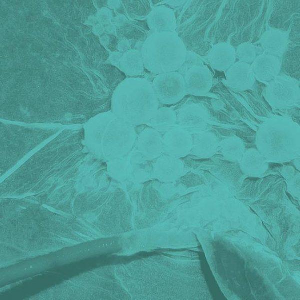
In addition to biochemical methods for exosome detection and quantitation, a number of biophysical techniques can be used for label-free exosome quantitation. Transmission electron microscopy (TEM), scanning electron microscopy (SEM), Cryo EM, and atomic force microscopy (AFM) have all been used to assess morphology and quality of exosome preparations, although cryo-EM studies call into question the cup-shape of exosomes as revealed by TEM.
Optical methods, such as NTA (Nanoparticle Tracking Analysis), provide accurate measurements of exosome size and concentration. NTA instruments are based on a conventional optical microscope and uses a laser light source to illuminate nano-scale particles within a sample introduced to the viewing unit with a disposable syringe. Enhanced by a near perfect black background, particles appear individually as point-scatterers moving under Brownian motion. Going one step further, Fluorescent NTA analysis can be performed to obtain a more accurate assessment of only the extracellular vesicles present in a heterogenous sample, as protein aggregates, membrane fractions, and other background particles will be excluded. SBI now offers NTA and Fluorescent NTA analysis via our custom services.
Conclusions
Whether you just need to know if you have exosomes present, or want a highly quantitative measurement of exosome amount, SBI is here to help. With a range of antibody- and activity-based options, a label-free analysis service using NTA, and expert technical support, we help you take full advantage of the power of exosomes.
If you have any questions about which method is the right one for your studies, don’t hesitate to contact the Technical Support Scientists here at SBI - they’re ready to help you learn the most you can about your exosomes.
REFERENCES
1. Savina, A., Vidal, M. & Colombo, M. I. The exosome pathway in K562 cells is regulated by Rab11. J. Cell Sci. 115, 2505–2515 (2002).
2. Gupta, S. & Knowlton, A. A. HSP60 trafficking in adult cardiac myocytes: role of the exosomal pathway. Am. J. Physiol. Heart Circ. Physiol. 292, H3052–3056 (2007).
