My Cart
You have no items in your shopping cart.
![]()
![]()
| Catalog Number | Description | Size | Price | Quantity | Add to Cart | |||
|---|---|---|---|---|---|---|---|---|
| EXOP-110A-1 | Frozen exosomes (>1x10^10) from HEK293 Human embryonic kidney cell line | 50 µg | $449 |
|
||||
| Catalog Number | Description | Size | Price | Quantity | Add to Cart | |||
|---|---|---|---|---|---|---|---|---|
| EXOP-110A-1 | Frozen exosomes (>1x10^10) from HEK293 Human embryonic kidney cell line | 50 µg | $449 |
|
||||
| Cat.# | |
|---|---|
| Exosomes isolated from cancer cell lines | |
| EXOP-100A-1 | MCF-7 Human breast cancer, noninvasive cell line |
| EXOP-105A-1 | MDA-MB-231 Human breast cancer, aggressive/invasive/metastatic cell line |
| EXOP-115A-1 | PC-3 Human prostate cancer cells derived from metastatic cancer cell line |
| EXOP-120A-1 | A549 Human non-small cell lung cancer cell line |
| EXOP-125A-1 | H841 Human small cell lung cancer cell line |
| EXOP-130A-1 | H196 Human small cell lung cancer cell line |
| EXOP-135A-1 | DMS114 Human small cell lung cancer cell line |
| Exosomes isolated from stem cell lines | |
| EXOP-140A-1 | PCS-500-011 Human pre-adipose derived mesenchymal stem cell |
| EXOP-145A-1 | PCS-500-012 Human bone marrow-derived mesenchymal stem cell line |
| Exosomes isolated from immune-related cell lines | |
| EXOP-150A-1 | BC-3 Human B lymphocyte cell line |
| EXOP-155A-1 | Jurkat Clone E6-1 Human T lymphocyte cell line |
| EXOP-160A-1 | JM1 Human T pre-B lymphoblast cell line |
| EXOP-165A-1 | RAW 264.7 Mouse macrophage cell line |
| EXOP-200A-1 | JAWS II Mouse bone marrow immature dendritic cell line |
| Exosomes isolated from general cell lines | |
| EXOP-110A-1 | HEK293 Human embryonic kidney cell line |
| XPAK100EX-G | XPack-GFP-loaded HEK293 exosomes |
| Exosomes isolated from biofluid exosomes | |
| EXOP-500A-1 | Human pooled serum (healthy donors) |
| EXOP-510A-1 | Human pooled saliva (healthy donors) |
| EXOP-520A-1 | Human pooled urine (healthy donors) |
| EXOP-530A-1 | Human pooled CSF (healthy donors) |
| EXOP-540A-1 | Human breast milk, normal (single donor) |
| EXOP-550A-1 | Human biofluids ascites fluid (single donor) |
| Exosome isolated from mouse | |
| EXOP-560M-1 | Mouse Pooled Serum |
Validated using Western blotting, NanoSight Analysis
All exosome preps are checked for the presence of CD63, a common exosome marker, via Western blotting (Figure 1).
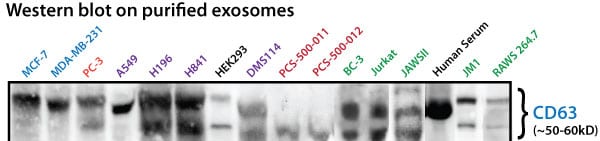
Figure 1. All exosome preps contain CD63. Aliquots of purified exosomes from the cell lines and from human serum were lysed with either RIPA or M-PER buffer to make exosome protein lysates. Approximately 20 ug of protein for each sample was separated on a gradient SDS-PAGE and then transferred to nitrocellulose membranes. The membranes were probed for CD63 profiles using SBI’s anti-CD63 antibody (cat# EXOAB-CD63A-1) at a 1:1,000 dilution. Bands were detected using the secondary HRP-conjugated antibody at 1:10,000 and blots imaged. All purified exosome preparations were positive, immunoreactive for CD63 with the expected, variable banding patterns common to published exosome CD63 profiles.
All exosome preps are also subjected to NanoSight Analysis to test for particle size and intactness (Figure 2).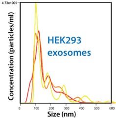
Figure 2. All exosome preps are checked for particle size and intactness via NanoSight. Approximately 5 µl of purified exosomes were added to 995 µl of 0.2 µm filtered 1X PBS (1:200 dilution). The diluted samples were incubated in a VWR 500 model ultrasonicator water bath set at 33°C for 10 minutes to ensure adequate exosome particle dispersion. The samples were diluted 1:10 then vortexed at 2.5k for 10 seconds. This eventual 1:2,000 dilution was used to gather between 1,000 to 3,000 particle tracks per sample analysis. The samples were then loaded into a NanoSight LM10HSB with a syringe pump and the sensitivity of the camera is set to auto 16 (the most sensitive auto-setting). All data were collected in triplicate. The purified exosomes displayed the expected size distribution profiles, with peak diameters from 90 – 110 nm and concentrations in the range expected for media exosomes at about 1 x 10¹⁰ exosomes/ml.
 Loading ... Loading ...