CMV-RFP-T2A-Luciferase Minicircle for In Vivo Imaging
- Foreign DNA-free—they are devoid of bacterial plasmid sequences, enabling gene therapy development and other in vivo applications
- More easily transfected—their small molecular size increases transfection efficiency
- Non-integrating—they reduce the risk of insertional mutagenesis due to their episomal nature
- Long-lasting—they deliver sustained expression over a period of weeks or months
- Safe—they are less toxic to cells, inducing lower stress and inflammation responses than other gene delivery systems
Products
| Catalog Number | Description | Size | Price | Quantity | Add to Cart | |||
|---|---|---|---|---|---|---|---|---|
| BLIV500MC-1 | Minicircle Dual Reporter: CMV-RFP-T2A-Luciferase DNA | 30 µg | $699 |
|
||||
| BLIV500MN-1 | Minicircle Dual Reporter: CMV-RFP-T2A-Luciferase Parental Plasmid | 10 µg | $969 |
|
||||
Overview
Overview
Clearly visualize cells in vivo and ex vivo for tracking cell fate and moreEasily label cell lines for tracking cell fate after implantation in an animal model or other imaging applications with SBI’s line of Bioluminescent, Fluorescent, and PET Imaging Vectors (BLIV). Available in a variety of promoter-reporter combinations that enable multiple imaging modalities—fluorescence with copGFP, bioluminescence with luciferase, and positron emission tomography (PET) with thymidine kinase (TK)—you can select from lentivector or minicircle technologies for both integrated and episomal expression.
SBI's minicricle-based imaging systems offer safe and efficient gene delivery with a number of benefits:
- Foreign DNA-free—they are devoid of bacterial plasmid sequences, enabling gene therapy development and other in vivo applications
- More easily transfected—their small molecular size increases transfection efficiency
- Non-integrating—they reduce the risk of insertional mutagenesis due to their episomal nature
- Long-lasting—they deliver sustained expression over a period of weeks or months
- Safe—they are less toxic to cells, inducing lower stress and inflammation responses than other gene delivery systems

The CMV-RFP-T2A-Luciferase Minicircle for In Vivo Imaging leverages SBI’s easy-to-use Minicircle Technology to deliver RFP and Luciferase to target cells. The reporters are co-expressed from a CMV promoter for high expression in most cell types, with co-expression mediated by a T2A element. The CMV-RFP-T2A-Luciferase Minicircle is available as both parental plasmid and ready-to-transfect minicircle DNA*.
Which BLIV construct should you choose?With a range of options, SBI’s vectors for in vivo imaging support a wide range of projects. Simply choose the vector that best fits your needs:
Choose a lentivector when:- You’re working with difficult-to-transfect cells
- You’d like to create stable reporter cell lines
- You’d like to integrate your reporter into the genome
- You’d like episomal expression
- You’d like to avoid introducing foreign sequences
| Promoter | Expression Level | Application |
|---|---|---|
| CMV | High | Commonly used in most cell lines (HeLa, HEK293, HT1080, etc.) |
| MSCV | High | Hematopoietic and stem cells |
| EF1α | Medium | Most cell types including primary cells and stem cells |
| PGK | Medium | Most cell types including primary cells and stem cells |
| UbC | Low | Most cell types including primary cells and stem cells |
References
How It Works
Supporting Data
Supporting Data
See SBI’s BLIV Vectors in action
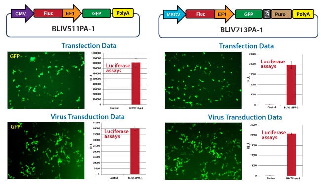
Figure 1. Examples of how SBI’s BLIV lentivectors deliver strong luciferase activity after both transduction and transfection.
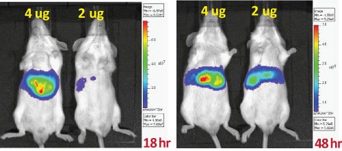
Figure 2. Examples of how SBI’s BLIV minicircle vectors deliver strong luciferase activity in small animal models.
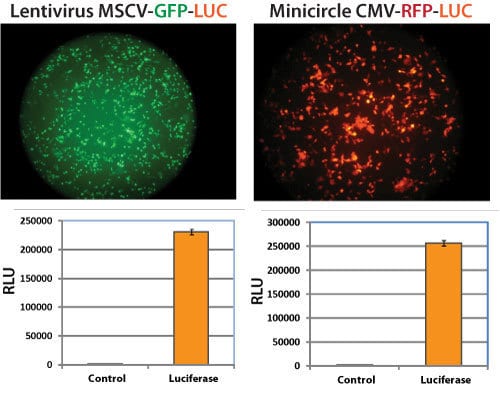
Figure 3. Examples of how SBI’s BLIV lentivectors and minicircle vectors can be used to generate stable cell lines with strong luciferase activity.
FAQs
Documentation
Citations
Related Products
Products
| Catalog Number | Description | Size | Price | Quantity | Add to Cart | |||
|---|---|---|---|---|---|---|---|---|
| BLIV500MC-1 | Minicircle Dual Reporter: CMV-RFP-T2A-Luciferase DNA | 30 µg | $699 |
|
||||
| BLIV500MN-1 | Minicircle Dual Reporter: CMV-RFP-T2A-Luciferase Parental Plasmid | 10 µg | $969 |
|
||||
Overview
Overview
Clearly visualize cells in vivo and ex vivo for tracking cell fate and moreEasily label cell lines for tracking cell fate after implantation in an animal model or other imaging applications with SBI’s line of Bioluminescent, Fluorescent, and PET Imaging Vectors (BLIV). Available in a variety of promoter-reporter combinations that enable multiple imaging modalities—fluorescence with copGFP, bioluminescence with luciferase, and positron emission tomography (PET) with thymidine kinase (TK)—you can select from lentivector or minicircle technologies for both integrated and episomal expression.
SBI's minicricle-based imaging systems offer safe and efficient gene delivery with a number of benefits:
- Foreign DNA-free—they are devoid of bacterial plasmid sequences, enabling gene therapy development and other in vivo applications
- More easily transfected—their small molecular size increases transfection efficiency
- Non-integrating—they reduce the risk of insertional mutagenesis due to their episomal nature
- Long-lasting—they deliver sustained expression over a period of weeks or months
- Safe—they are less toxic to cells, inducing lower stress and inflammation responses than other gene delivery systems

The CMV-RFP-T2A-Luciferase Minicircle for In Vivo Imaging leverages SBI’s easy-to-use Minicircle Technology to deliver RFP and Luciferase to target cells. The reporters are co-expressed from a CMV promoter for high expression in most cell types, with co-expression mediated by a T2A element. The CMV-RFP-T2A-Luciferase Minicircle is available as both parental plasmid and ready-to-transfect minicircle DNA*.
Which BLIV construct should you choose?With a range of options, SBI’s vectors for in vivo imaging support a wide range of projects. Simply choose the vector that best fits your needs:
Choose a lentivector when:- You’re working with difficult-to-transfect cells
- You’d like to create stable reporter cell lines
- You’d like to integrate your reporter into the genome
- You’d like episomal expression
- You’d like to avoid introducing foreign sequences
| Promoter | Expression Level | Application |
|---|---|---|
| CMV | High | Commonly used in most cell lines (HeLa, HEK293, HT1080, etc.) |
| MSCV | High | Hematopoietic and stem cells |
| EF1α | Medium | Most cell types including primary cells and stem cells |
| PGK | Medium | Most cell types including primary cells and stem cells |
| UbC | Low | Most cell types including primary cells and stem cells |
References
How It Works
Supporting Data
Supporting Data
See SBI’s BLIV Vectors in action
Figure 1. Examples of how SBI’s BLIV lentivectors deliver strong luciferase activity after both transduction and transfection.
Figure 2. Examples of how SBI’s BLIV minicircle vectors deliver strong luciferase activity in small animal models.
Figure 3. Examples of how SBI’s BLIV lentivectors and minicircle vectors can be used to generate stable cell lines with strong luciferase activity.


